


Fitness Metrics Profile + Thyroid Profile is designed to run the following 20 tests accurately:
- Estradiol (Estrogen) (E2),
- Progesterone (Pg),
- Testosterone (T),
- DHEA-S (DS),
- Sex Hormone Binding Globulin (SHBG),
- Cortisol (C),
- Thyroid Stimulating Hormone (TSH),
- Vitamin D2,
- Vitamin D3,
- Triglycerides (TG),
- Total Cholesterol (CH),
- HDL Cholesterol (HDL),
- LDL Cholesterol (LDL)
Add on Thyroid Profile:
- Free Triiodothyronine (fT3),
- Free Thyroxine (fT4) and
- Thyroid Peroxidase Antibody (TPOab)
- Hemoglobin A1c (HbA1c)
- High Sensitivity C-Reactive Protein (hs-CRP)
- Luteinizing (Lh)
Test Result: You will receive your test result 3-5 working days after the laboratory receives your sample. You will see your hormone levels in graphics and numbers on your test results. In the comments, you will also see laboratory comments by a hormone specialist PhD. You will find a doctor's analysis of your hormone levels and what to do next.
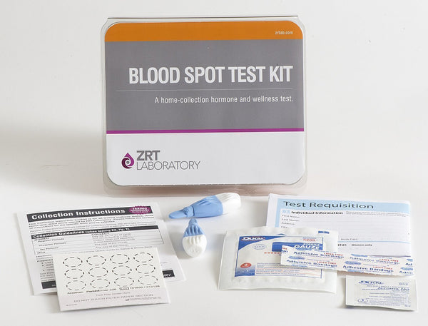
- Click to see >> Fitness Metrics Sample Collection Instruction
- Collect samples from the comfort of your home and post them to our lab.
- The test is suitable for both adults and children
- The test must be used within 12 months after the purchase date.
- The test kit includes a laboratory fee: no additional laboratory cost or tax.
- Customers are responsible for shipping their samples to the laboratory.
This test kit will give you an understanding of your hormone levels and determine your vitamin D status and blood lipid levels. By understanding your hormones, lipids, and vitamin D status and tracking them over time, you can monitor how fitness training affects your overall health and prevent overtraining syndrome.
The Fitness Metrics Profile
The Fitness Metrics Profile will give you a basic understanding of your hormone levels and determine your vitamin D status and blood lipid levels. By understanding your hormones, lipids, and vitamin D status and tracking them over time, you can monitor how fitness training affects your overall health and prevent overtraining syndrome.
The optional add-ons give a fuller picture of overall health and well-being, including a complete picture of the thyroid, showing where thyroid imbalance can influence workouts. Knowing LH levels helps us understand the effects of exercise on how the body produces endogenous steroids. The optional cardiometabolic insulin, HbA1c and hs-CRP allow you to track how increased fitness activity positively impacts your health.
Who Benefits from Fitness Profile Testing?
People who are:
- Looking to start a fitness program
- Interested in losing weight
- Monitoring fitness or weight loss goals
Estradiol (E2) (Estrogen - Oestrogen)
Estradiol (Estrogen) is at optimal physiological levels in women and promotes a healthy distribution of fat in hips, thighs, breasts, and under the skin (subcutaneously). However, in excess and without progesterone, oestrogen predisposes to unhealthy surplus weight gain in these tissues. Men generally have much lower levels of estradiol and higher testosterone, responsible for greater muscle mass and less fat distribution in body areas normally seen in women. In overweight men, testosterone levels drop, and oestrogens rise, leading to the same problematic weight gain in the hips, thighs, and breasts (referred to as gynecomastia) as seen in women.
Progesterone (Pg)
Progesterone, in addition to keeping oestrogen levels in check, aids weight management by supporting thyroid metabolism, helping the body use and eliminate fats, and acting as a natural diuretic. Progesterone and oestrogen help control how insulin is released and body fat is stored in the proper ratios. In addition, as the precursor of cortisol, progesterone supports adrenal regulation of blood glucose, while its natural calming properties may relieve stress-related overeating and food cravings.
Testosterone (T) And DHEAS (DS)
Testosterone and DHEA-S (DS) are androgens that increase lean muscle mass and metabolic rate. As androgen levels decline, muscle mass also decreases with a corresponding increase in adiposity. Low androgens can also reduce vitality and tolerance for exercise. With its resulting hormone imbalances, weight gain can trigger a drop in testosterone in men. The aromatase enzyme within fat tissue converts androgens to oestrogens, contributing to a female-type body fat distribution, including breast tissue development. In women with polycystic ovarian syndrome (PCOS), high testosterone and DHEA are linked to insulin resistance and weight gain, particularly in the abdomen.
Cortisol (C)
Imbalances can create problems with blood sugar control, sleep patterns, appetite, food cravings, and exercise tolerance. Under stress, excessive cortisol production, particularly in concert with insulin, promotes fat storage in abdominal adipose tissue. This visceral fat is closely associated with insulin resistance and metabolic syndrome and is thus more hazardous to health. In addition, chronically elevated cortisol is a known risk factor for pre-diabetes and cardiovascular disease.
Thyroid Stimulating Hormone (TSH)
Even within the high-normal range, TSH elevations are linked with hypothyroidism, low metabolic rate and obesity. Hypothyroidism is linked to elevated cortisol and can also be a consequence of oral oestrogen therapy, which increases the production of binding proteins that reduce thyroid hormone bioavailability.
Vitamin D (D2, D3)
Vitamin D deficiency is common in obesity and is particularly associated with hyperinsulinemia and visceral fat. Whether by cause or effect, identifying and correcting vitamin D2 and D3 deficiency may improve insulin sensitivity.
Most people are familiar with vitamin D's role in preventing children's rickets and helping the body absorb calcium from the diet.
Recently, research has shown that vitamin D is vital in protecting the body from a wide range of diseases. Disorders linked with vitamin D deficiency include:
- Stroke
- Flu
- Cardiovascular disease
- Osteoporosis
- Osteomalacia
- Several forms of cancer
- Some autoimmune diseases, such as multiple Sclerosis
- Rheumatoid Arthritis
- Type I Diabetes & Type II Diabetes
- Breast and Colon Cancer (linked to Vitamin D Deficiency)
- Depression and even schizophrenia
Vitamin D is a prohormone and not technically a vitamin: a vitamin is defined as a substance not made naturally by the body but must be supplied in the diet to maintain life processes. But in fact, we make most of our vitamin D by the action of ultraviolet light (sunlight) on the vitamin D originator found in our skin. As a result, we only get minimal amounts of vitamin D from our diet. However, increasingly, it is added to foods eaten by children in an attempt to prevent rickets in the population.
Total Cholesterol (CH)
The body requires cholesterol as a precursor to steroid hormone synthesis and as a component of cell membranes. However, in excessive amounts, it is a vital component of coronary heart disease risk because of its contribution to coronary atherosclerosis. Atherosclerotic plaque is primarily composed of cholesterol, as with other risk factors; high blood cholesterol levels are more significant when other cardiometabolic parameters are abnormal or in patients with diabetes or cardiovascular disease.
Triglycerides (TG)
Triglycerides enter the circulation as the end-product of digesting dietary fat; the liver also synthesizes them. They are an essential energy source for the body and are stored in fat cells. Elevated blood levels, or hypertriglyceridemia, often found in untreated diabetes and obesity, are an established indicator of atherogenic dyslipidemia. The National Cholesterol Education Program defines fasting triglyceride levels of 150 mg/dL or above as one of the diagnostic criteria for metabolic syndrome. However, some studies have shown that fasting levels lower than 100 mg/dL should be considered as a more optimal cutoff in coronary heart disease risk assessment. The inflammatory state leading to the development of atherosclerosis may be triggered by "postprandial dysmetabolism," a condition characterized by unusually high levels of glucose and triglycerides after a meal. Postprandial hypertriglyceridemia indicates the presence of remnant lipoproteins, which are believed to promote atherosclerosis and are also linked with insulin resistance and obesity. Several studies have found that triglyceride levels measured 2-4 hours after meals highly predict cardiovascular events, especially in women. Nonfasting levels >200 mg/dL are suggestive of postprandial dysmetabolism.
HDL Cholesterol (HDL)
Cholesterol bound to high-density lipoprotein (HDL) in the blood is known as HDL cholesterol. A low level of circulating HDL cholesterol is one of the established criteria for diagnosing metabolic syndrome and has long been regarded as a powerful predictor of cardiovascular disease in diabetics and non-diabetics. In a large cohort from the Framingham Study, a high total cholesterol/HDL cholesterol or LDL cholesterol/HDL cholesterol ratio was associated with increased coronary heart disease risk, and a high HDL cholesterol level was associated with reduced risk in both men and women. Currently, the LDL cholesterol/HDL cholesterol ratio is regarded as a reliable tool for evaluating cardiovascular disease risk. While absolute values of each are still considered by the National Cholesterol Education Program (NCEP) as the optimal diagnostic indicators, the ratios that doctors and researchers currently accept are as follows: total cholesterol: HDL cholesterol ratio – optimally below 4; LDL cholesterol: HDL cholesterol ratio – optimally below 3. The current NCEP recommendation to reduce the risk of cardiovascular disease is to maintain an HDL cholesterol level >40 mg/dL in both men and women.
Sex Hormone Binding Globulin (SHBG)
SHBG binds and transports both testosterone and estrogens in the bloodstream and, therefore, regulates the relative amounts of free and bound hormones and, consequently, their bioavailability to target tissues. SHBG is a protein produced by the liver in response to exposure to any estrogen. Testosterone binds about three times more tightly to SHBG than estradiol, so this increase in SHBG due to estrogen exposure causes the relative proportion of bioavailable testosterone to estradiol to decrease even further, exacerbating the symptoms of testosterone deficiency. Many factors, in addition to estrogen exposure, can affect SHBG levels. For example, thyroid hormone increases SHBG production, whereas insulin, on the other hand, decreases SHBG levels. In young men, testosterone levels are usually high and SHBG low, making most of the testosterone bioavailable. However, as men age, gain weight, and their estrogen levels increase, SHBG also rises, decreasing bioavailable testosterone. Measuring SHBG in the blood indicates the overall exposure to estrogens and the bioavailable (free) fraction of testosterone (calculated from the ratio of testosterone to SHBG).
**ADD ON THYROID PROFILE
Free T3 – Triiodothyronine
The active thyroid hormone regulates the metabolic activity of cells. Free T3 is the non-protein-bound fraction circulating in the blood, representing about 0.4% of the total circulating T3 available to tissues. Therefore, elevated T3 levels are seen in hyperthyroid patients, but levels can be normal in hypothyroid patients because it does not represent the intracellular conversion of T4 to T3, which comprises about 60% of all T3 formed in tissues.
TPO – Thyroid Peroxidase Antibodies
Thyroid peroxidase is an enzyme used by the thyroid gland to manufacture thyroid hormones by liberating iodine for attachment to tyrosine residues on thyroglobulin. In patients with autoimmune thyroiditis (predominantly Hashimoto's disease), the body produces antibodies that attack the thyroid gland, and levels of these antibodies in the blood can diagnose this condition and indicate the extent of the disease.
Free T4- Thyroxine
The predominant hormone is produced by the thyroid gland. It is an inactive hormone and is converted to its active form, T3, within cells. Free T4 is the non-protein bound fraction of the T4 circulating in the blood, representing about 0.04% of the total circulating T4 available to tissues. Low TSH combined with low T4 levels indicates hyperthyroidism. High TSH and low T4 abdicate a thyroid gland disease, such as autoimmune thyroiditis (Hashimoto's).
Hemoglobin A1c (HbA1c)
Haemoglobin A1c (HbA1c) is a glycated form of haemoglobin that results from the binding of haemoglobin in red blood cells to glucose in the bloodstream. Once the haemoglobin has bound to glucose, it remains glycated. Circulating red blood cells have a lifespan of 120 days; therefore, the amount of HbA1c at any time reflects the average exposure of red blood cells to glucose over the previous three months. It can consequently indicate impaired glucose tolerance even when occasional fasting plasma glucose measurements are normal.
High Sensitivity C-Reactive Protein (hs-CRP)
C-reactive protein (CRP) is an established marker of inflammation and has recently been suggested to be an important contributor to the pro-inflammatory and prothrombotic elements of cardiovascular disease (CVD) risk.
Extremely high CRP levels are seen in acute inflammatory states, but the small elevations indicative of the pro-inflammatory and prothrombotic states implicated in the metabolic syndrome require high sensitivity assays and are thus referred to as hs-CRP levels.
Studies have shown correlations between elevated hs-CRP and increased risk of future heart attacks, ischemic stroke, and peripheral arterial disease. Overweight, obese, insulin-resistant, and diabetic individuals typically have elevated hs-CRP levels; elevations in hs-CRP levels have also been found to predict the development of diabetes—lifestyle changes such as aerobic exercise, weight loss, and smoking cessation lower hs-CRP levels. Levels below 3.0 mg/L are considered normal; 3.1—10 mg/L is elevated in the context of CVD risk, and above 10 mg/ L is very high, more likely indicating an acute inflammatory event due to infection or trauma.
Luteinizing hormone (LH)
Luteinizing hormone (LH), a glycoprotein hormone produced by the anterior pituitary, is essential for reproduction in both men and women.
In women, controlled by a negative feedback loop involving several ovarian hormones, gonadotropin-releasing hormone (GnRH) is secreted in pulses from the hypothalamus, which stimulates LH production from the pituitary gland. In a normal menstrual cycle, a surge of LH production lasting around 48 hours occurs at the end of the follicular phase. This sudden burst of LH causes luteinization of the ovarian follicles and triggers ovulation.
In men, LH acts on the Leydig cells of the testes to stimulate the production of testosterone, which is necessary for sexual function and spermatogenesis. LH levels are helpful for the clinical assessment of infertility: low levels in men can result in hypogonadism and insufficient sperm production, while in women, LH levels are used to determine the occurrence of ovulation for couples trying to conceive. High LH levels are seen in polycystic ovarian syndrome and in precocious puberty; levels are similar to those seen in reproductive-age individuals instead of the lower levels normally seen in children.
LH levels can also be used in diagnosing pathologies of the hypothalamus or pituitary. As women enter menopause, LH levels rise as ovarian hormone production declines, reducing the negative feedback on GnRH production. LH testing can help evaluate a woman's menopausal status. Ranges for blood spot LH in premenopausal women (luteal phase) are 0.5—12.8 U/ L, in premenopausal women (follicular phase) 1.6—9.3 U/L, in postmenopausal women 15—64 U/L, and in men 1.0—8.4 U/L
How to Use