Female Hormone Test (Full Profile) is designed to measure 28 following essential hormones and elements accurately:
- Estradiol (Estrogen) (E2),
- Progesterone (Pg),
- Testosterone (T),
- DHEA-S (DS),
- Sex Hormone Binding Globulin (SHBG),
- Cortisol (C),
- Thyroid Stimulating Hormone (TSH),
- Free Triiodothyronine (fT3),
- Free Thyroxine (fT4),
- Thyroid Peroxidase Antibody (TPO),
- High Sensitivity C-Reactive Protein (hs-CRP),
- Thyroglobulin (Tgbn),
- Luteinising(Lh),
- Thyroxine (t4),
- Vitamin D 25-oh total Vitamin D2,
- Vitamin D 25-oh total Vitamin D3,
- Cadmium (Cd),
- Mercury (Hg),
- Selenium (Se),
- Zinc (Zn),
- Magnesium (Mg),
- Copper (Cu),
- Insulin (IN),
- High Sensitivity C-Reactive Protein (hs-CRP),
- Haemoglobin (HbA1c),
- Triglycerides (TG),
- Total Cholesterol (CH),
- HDL Cholesterol (HDL).
Test Result: You will receive your test result 3-5 working days after the laboratory receives your sample. You will see your hormone levels in graphics and numbers on your test results. You will also see laboratory comments by a hormone specialist PhD in the comments. You will find a doctor's analysis of your hormone levels and what to do next.
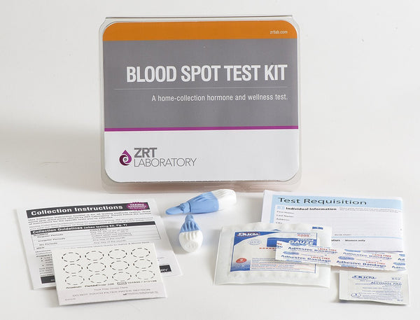
- Collect samples from the comfort of your home and post them to our lab.
- The test must be used within 12 months after the purchase date.
- The test kit includes a laboratory fee: no additional laboratory cost or tax.
- Customers are responsible for shipping their samples to the laboratory.
- Click the link to see the Sample Test Result Report in Blood Spot
Having too much or too little of a particular hormone in the body can cause hormonal imbalance.
In addition, a hormonal imbalance can lead to irregular periods and affect fertility and the ability to conceive.
Understanding your hormone levels, how they fluctuate over your cycle and how they change over different life stages can help you keep track of any imbalances that may make you experience some of the symptoms below.
Symptoms related to Hormonal Imbalance in Women include:
- Burned Out Feeling
- Hot Flashes
- Decreased Sweating
- Cold Body Temperature
- Decreased Stamina
- Decreased Flexibility
- Slow Pulse Rate
- Memory Lapses
- Sleep Disturbances
- Poor Concentration
- Dizzy spells
- Mood Swings
- Decreased Mental Sharpness
- Nervous
- Headaches
- Depressed
- Apathy
- Anxious
- Stress
- Aggressive Behaviours
- Irritable
- Difficulty Sleeping
- Mental Fatigue
- Morning Fatigue
- Increased Forgetfulness
- Evening Fatigue
- Tearful
- Allergies
- Sensitivity to Chemicals
- Elevated Triglyceride
- Breast Cancer
- Fibrocystic Breasts
- Decreased Urine Flow
- Increased Urinary Urged
- Incontinence
- Bone Loss
- Decreased Muscle Size
- Swelling or Puffy Eyes/face
- Nails Breaking or Brittle
- Rapid Heartbeat
- Low Blood Sugar
The Hormones Tested in Our Female Profile and Why
Estradiol (Estrogen) and progesterone hormone imbalance:
Levels and their ratio are an index of estrogen/progesterone balance. An excess of estradiol, relative to progesterone, can explain many symptoms in reproductive-age women, including endometrial hyperplasia, pre-menstrual syndrome, fibrocystic breasts, and uterine fibroids. In older women using estrogen supplements alone, a progesterone deficiency can also result in estrogen dominance symptoms, including weight gain in the hips and thighs, fibrocystic and tender breasts, uterine fibroids, irritability, water retention, and thyroid problems. These symptoms are also seen in some women approaching menopause, whose estrogen levels swing wildly from high to low without the balancing effects of progesterone. If estrogen dominance is not corrected, it can lead to cancers of the uterus and breasts and insulin resistance. With the onset of menopause, when ovarian estrogen and progesterone production declines, a new subset of symptoms can result from low estradiol levels, including hot flashes, night sweats, vaginal dryness, sleep disturbances, foggy thinking, more rapid skin ageing, and bone loss. Therefore, maintaining appropriate levels of estradiol, adequately balanced with progesterone, at any age is essential for optimal health.
Testosterone and Sex Hormone Binding Globulin (SHBG) hormone imbalance:
Testosterone levels can also be either too high or too low. Testosterone in excess, often caused by ovarian cysts, leads to conditions such as excessive facial and body hair, acne, and oily skin and hair. Polycystic ovarian syndrome (PCOS) is thought to be caused, in part, by insulin resistance. On the other hand, too little testosterone is often caused by excessive stress, medications, contraceptives, and surgical removal of the ovaries. This leads to symptoms of androgen deficiency including loss of libido, thinning skin, vaginal dryness, loss of bone and muscle mass, depression, and memory lapses. SHBG is a protein produced by the liver in response to exposure to any type of estrogen, whether produced naturally by the body, consumed as a synthetic oral contraceptive estrogen, estrogen therapy, or as foods or herbs (phytoestrogens). Released from the liver into the bloodstream, SHBG binds tightly to circulating estradiol and testosterone, preventing their rapid metabolism and clearance and limiting their bioavailability to tissues. SHBG gives a good index of the extent of the body’s overall exposure to estrogens. The SHBG level is also used to calculate free (unbound) testosterone levels when blood spot is used instead of saliva to measure sex hormones.
DHEA Hormone Imbalance:
Mostly found in the circulation in its conjugated form, DHEA sulfate (DHEA-S), is a hormone produced by the adrenal glands, and levels generally reflect adrenal gland function. It is a precursor for the production of estrogens and testosterone and is therefore normally present in greater quantities than all the other steroid hormones. Its production is highest in the late teens to early 20s and declines gradually with age. Like cortisol, it is involved with immune function, and a balance between the two is essential. Low DHEA can result in reduced libido and general malaise, while high DHEA can have masculinizing effects on women because it metabolizes androgens, including testosterone. Because of its conversion to estrogens and androgens, it is important to monitor levels of these hormones, as well as levels of DHEA, during supplementation.
Cortisol hormonal imbalance is an indicator of adrenal function and exposure to stressors. Under normal circumstances, adrenal cortisol production shows a diurnal variation and is highest early in the morning, soon after waking, falling to lower levels in the evening. Normal cortisol production shows a healthy ability to respond to stress. Low cortisol levels can indicate adrenal fatigue (a reduced ability to respond to stressors) and can leave the body more vulnerable to poor blood sugar regulation and immune system dysfunction. Chronically high cortisol is a consequence of high, constant exposure to stressors, and this has serious implications for long-term health, including an increased risk of cancer, osteoporosis, and possibly Alzheimer’s disease.
Free T4, free T3, TSH, and TPO hormone imbalance tests can indicate the presence of an imbalance in thyroid function, which can cause a wide variety of symptoms, including feeling cold all the time, low stamina, fatigue (particularly in the evening), depression, low sex drive, weight gain, and high cholesterol. Thyroid deficiency can also be a cause of infertility, which is why these tests are included in the Female Fertility Profiles.
LH hormone imbalance tests are included in the Female Fertility Profile to give information on the possible presence of PCOS (elevated LH/FSH).
TSH – Thyroid Stimulating Hormone
Produced by the Pituitary. TSH acts on the thyroid gland to stimulate the production of the thyroid hormones T4 and T3. Higher than normal TSH can indicate overproduction of or excessive supplementation with T4 and/or T3, Which acts in negative feedback on the pituitary to reduce TSH production. Low TSH can also be caused by problems in the pituitary gland itself, which result in insufficient TSH being produced to stimulate the thyroid (secondary hypothyroidism).
Free T4- Thyroxine
The predominant hormone is produced by the thyroid gland. It is an inactive hormone and is converted to its active form, T3, within cells. Free T4 is the non-protein bound fraction of the T4 circulating in the blood, representing about 0.04% of the total circulating T4, which is available to tissues. Low TSH combined with low T4 levels indicates hyperthyroidism. High TSH and low T4 abdicate a thyroid gland disease, such as autoimmune thyroiditis (Hashimoto’s).
Total T4 – Thyroxine
Total T4 includes both free T4 and protein-bound T4 and therefore represents the thyroid gland’s capacity to synthesise, process, and release T4 into the bloodstream. In contrast, free T4 represents only the circulating hormone that is bioavailable and not tightly complex with thyroid-binding globulin (TBG). Certain conditions, like oral estrogen usage or pregnancy, can
cause total levels to change due to liver induction of TBG. This can result in no change in free T4 or lower bioavailable levels of free T4 even though total T4 increases.
Free T3 – Triiodothyronine
The active thyroid hormone regulates the metabolic activity of cells. Free T3 is the non-protein-bound fraction circulating in the blood, representing about 0.4% of the total circulating T3, which is available to tissues. Elevated T3 levels are seen in hyperthyroid patients, but levels can be normal in hypothyroid patients because it does not represent the intracellular conversion of T4 to T3, which comprises about 60% of all T3 formed in tissues.
TPO – Thyroid Peroxidase Antibodies
Thyroid peroxidase is an enzyme used by the thyroid gland in the manufacture of thyroid hormones by liberating iodine for attachment to tyrosine residues on thyroglobulin. In patients with autoimmune thyroiditis (predominantly Hashimoto’s disease), the body produces antibodies that attack the thyroid gland, and levels of these antibodies in the blood can diagnose this condition and indicate the extent of the disease.
Thyroglobulin
A protein that is rich in tyrosine and synthesised only in the thyroid gland. When bound to iodine, tyrosine residues in thyroglobulin become the source material for the synthesis of the thyroid hormones T3 and T4. When iodine levels are low, high levels of thyroglobulin can be found in the blood as iodine-poor thyroglobulin builds up and leaks from the thyroid into the bloodstream. Levels of thyroglobulin are an indicator of a person’s average iodine exposure over a period of weeks: the
greater the iodine exposure, the lower the thyroglobulin level. Elevated thyroglobulin, in the absence of more serious thyroid diseases such as thyroid cancer, which results in very high blood thyroglobulin levels, indicates low iodine status.
Hemoglobin A1c (HbA1c)
It is a glycated form of haemoglobin that results from the binding of haemoglobin in red blood cells to glucose in the bloodstream. Once the haemoglobin has bound to glucose, it remains glycated. Circulating red blood cells have a lifespan of 120 days, therefore the amount of HbA1c at any time point reflects the average exposure of red blood cells to glucose over the previous 3 months. It can, therefore, indicate impaired glucose tolerance even when occasional fasting plasma glucose measurements are normal.
High Sensitivity C-Reactive Protein (hs-CRP)
C-reactive protein (CRP) is an established marker of inflammation and has recently been suggested to be an important contributor to the pro-inflammatory and prothrombotic elements of cardiovascular disease (CVD) risk.
Extremely high CRP levels are seen in acute inflammatory states, but the small elevations that are indicative of the pro-inflammatory and pro-thrombotic states implicated in the metabolic syndrome require high sensitivity assays and are thus referred to as hs-CRP levels.
Studies have shown correlations between elevated hs-CRP and increased risk of future heart attacks, ischemic stroke, and peripheral arterial disease. Overweight, obese, insulin-resistant, and diabetic individuals typically have elevated hs-CRP levels; elevations in hs-CRP levels have also been found to predict the development of diabetes. Lifestyle changes such as aerobic exercise, weight loss, and smoking cessation lower hs-CRP levels. Levels below 3.0 mg/L are considered to be normal; 3.1—10 mg/L is elevated in the context of CVD risk, and above 10 mg/ L is very high, more likely indicating an acute inflammatory event due to infection or trauma.
Vitamin D in Blood Spot
Vitamin D Deficiency – A Real Problem
Most people are familiar with vitamin D’s role in preventing rickets in children and in helping the body absorb calcium from the diet.
Recently, research has shown that vitamin D is important in protecting the body from a wide range of diseases. Disorders linked with vitamin D deficiency include:
- Stroke
- Flu
- Cardiovascular disease
- Osteoporosis
- Osteomalacia
- Several forms of cancer,
- Some autoimmune diseases, such as multiple Sclerosis
- Rheumatoid Arthritis
- Type I Diabetes & Type II Diabetes
- Breast and Colon Cancer (linked to Vitamin D Deficiency)
- Depression and even schizophrenia.
Vitamin D is a prohormone and not technically a vitamin: a vitamin is defined as a substance that is not made naturally by the body but must be supplied in the diet to maintain life processes. But in fact, we make most of our vitamin D by the action of ultraviolet light (sunlight) on the vitamin D originator found in our skin. As a result, we only get very small amounts of vitamin D from our diet, although increasingly, it is added to foods eaten by children to prevent rickets in the population.
Cause of Vitamin D Deficiency
A major cause of deficiency is not getting enough sun.
This happens because people don’t spend much time outdoors, women in particular often have much of their skin area covered for cultural reasons, and the use of sunscreen also blocks the formation of vitamin D in the skin.
Why Vitamin D Deficiency Test?
Testing for vitamin D is, therefore, an important screening test, especially if you spend much of your time indoors or live in a colder climate. This blood spot test measures both the natural form of Vitamin D (D3) as well as D2, the form that is used in many supplements. So, testing can be used to monitor vitamin D supplementation to ensure you are getting the right amount for optimum health.
How to Use